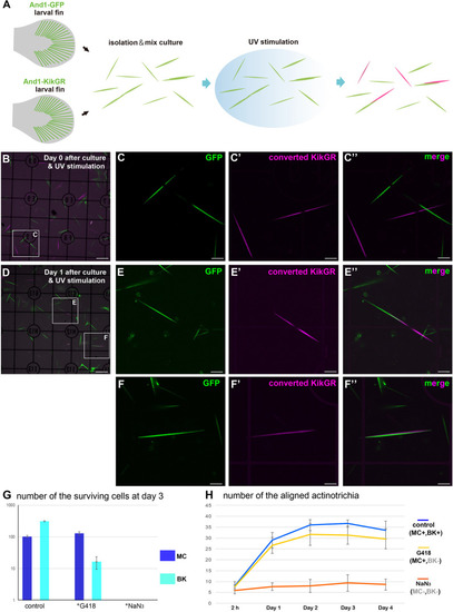
Mesenchymal cells are essential for the in vitro alignment of actinotrichia. (A) Illustration of the procedure for the in vitro mix culture experiment. Actinotrichia and cells were harvested from two different TG larval fins (TG; and1 1.4k: And1-GFP, and1 1.4k: And1-KikGR) and cultured on a Matrigel-coated dish. The isolated actinotrichia were stimulated by UV radiation in vitro and the aligned actinotrichia composed of fibrils labeled by two different fluorescent proteins were counted. (B) The fluorescence image at Day 0 (after 2 h of culture and UV stimulation). (C–C′′) Magnified images of the white box in (B). (D) The fluorescence image at Day 1 after culture and UV stimulation. (E–E′′) Magnified images of the white box in (D). Two actinotrichia were connected tip-to-tip and their orientation was aligned. (F–F′′) Magnified images of the white box in (D). Two actinotrichia were connected with side-to-side and their orientation was aligned. (G) Number of the surviving mesenchymal cells and basal keratinocytes under the control, G418 (200 μg/ml)-treated condition and NaN3 (0.1%)-treated condition. (H) The aligned actinotrichia were increased in the G418-treated condition (only mesenchymal cells alive) but not in the NaN3-treated condition (cell-free state). MC, mesenchymal cell; BK, basal keratinocyte. Scale bars: 100 μm in (B,D), 20 μm in (C-C′′,E-E′′,F-F′′).
|
