FIGURE

FIGURE 4

ID
ZDB-FIG-201121-40
Publication
Kuroda et al., 2020 - The Physical Role of Mesenchymal Cells Driven by the Actin Cytoskeleton Is Essential for the Orientation of Collagen Fibrils in Zebrafish Fins
Other Figures
All Figure Page
Back to All Figure Page
FIGURE 4

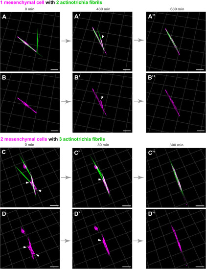
Live cell imaging of cultured mesenchymal cells holding the actinotrichia fibrils. Captured time-lapse images for the interaction between mesenchymal cells and actinotrichia fibrils. Mesenchymal cells and actinotrichia were isolated from the fins of F1 TG larvae [TG; 5xand1(MC): Lifeact-mCherry/5xand1(2P): And1full-GFP] and cultured on a Matrigel-coated dish. (A) A single mesenchymal cell initially surrounded one of two different actinotrichia fibrils. (A′) At 430 min, the mesenchymal cell started to develop a filopodia like structure (white arrowheads) and to hold another fibril. Finally, (A′′) at 630 min, two fibrils were completely aligned in the direction of the longitudinal axis of the mesenchymal cell. The fluorescence images of only a mesenchymal cell are shown in the bottom panels (B–B′′). (C) Two mesenchymal cells initially developed their pseudopodia (white arrowheads) and attached to three different fibrils, and (C′) gradually changed their morphology and moved the fibrils. (C′′) At 300 min, the two cells extended in the same direction and all of three fibrils were oriented along the longitudinal axis of the two mesenchymal cells. The fluorescence images of only two mesenchymal cells are shown in bottom panels (D–D′′). Scale bars: 20 μm.

Expression Data

Expression Detail
Antibody Labeling
Phenotype Data

Phenotype Detail
Acknowledgments
This image is the copyrighted work of the attributed author or publisher, and ZFIN has permission only to display this image to its users. Additional permissions should be obtained from the applicable author or publisher of the image. Full text @ Front Cell Dev Biol