FIGURE

Figure 4—figure supplement 1.

ID
ZDB-FIG-200822-22
Publication
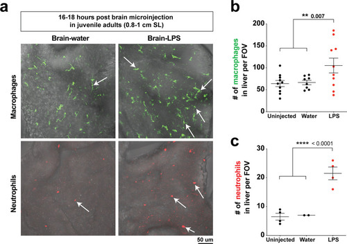
Yang et al., 2020 - Drainage of inflammatory macromolecules from brain to periphery targets the liver for macrophage infiltration
Other Figures
All Figure Page
Back to All Figure Page
Figure 4—figure supplement 1.

Hepatic response to brain-LPS challenge still evident after establishment of Kupffer cells and adult vasculature/brain structures in juvenile adults.

(a) Live ex vivo imaging of dissected liver from juvenile adult zebrafish after microinjection of either water vehicle or LPS in the brain at 16–18 hpi. Top, arrows point to macrophages in liver using the mpeg1:GFP transgene. Bottom, arrows point to neutrophils in the liver using the lyz:mCherry transgene. (b) Quantification of macrophage density (# mpeg1:GFP+ cell per field of view, FOV). (c) Quantification of neutrophil density (# lyz:mCherry+ cell per FOV). Scatter plots show uninjected controls, or animals microinjected with water vehicle or LPS in the brain. Each field of view (FOV) equals 0.045 squared mm. Statistical significance was determined by a two-tailed Student’s t-test.

Expression Data

Expression Detail
Antibody Labeling
Phenotype Data

Phenotype Detail
Acknowledgments
This image is the copyrighted work of the attributed author or publisher, and ZFIN has permission only to display this image to its users. Additional permissions should be obtained from the applicable author or publisher of the image. Full text @ Elife