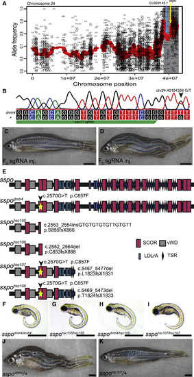
Functional Genetic Studies Link Scospondin with dmh4 Mutant Phenotypes (A) dmh4 mutation mapping graph for chromosome 24. Region of linkage (gray box) with locations of CU929145.1 (blue arrow) and sspo (yellow arrow) indicated. (B) Schematic depicting position and sequence of sspo c.2570G > T variant and CRISPR exon 17 sgRNA. (C and D) Following embryonic injection of Cas9 and sspo exon 17 sgRNA, most F0 fish with mosaic sspo mutations appear normal (C), whereas some develop spinal curvatures (n = 6/181; D). Scale bars represent 2 mm. (E) Schematic representation of the location and predicted functional outcomes for sspo mutations reported in this paper. SCORs, SCO-spondin repeat domains; TSR, thrombospondin type 1 repeat; vWDs, von Willebrand domains; LDLrA, low density lipoprotein receptor type A domain. (F–I) Lateral views of sspo mutant embryos at 3 dpf, demonstrating CTD phenotypes associated with dmh4/dmh4 (F), hsc105/hsc105 (G), dmh4/hsc105 (H), and hsc107/hsc107 (I) allelic combinations. Scale bars represent 1 mm. (J) Representative photo of an adult sspodmh4/+ zebrafish with obvious scoliosis. Scale bar represents 2 mm. (K) Representative photo of an adult sspohcs107/+ zebrafish, demonstrating that sspo loss-of-function mutations introduced in cis with the dmh4 c.2570G > T variant can suppress dominant scoliosis phenotypes. Scale bar represents 2 mm. See also Figure S2.
|
