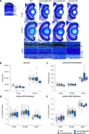
Morphologic analysis of cacna2d4-KO retinae. (A) Semithin plastic sections (3 μm) of WT, cacna2d4a-KO, cacna2d4b-KO and double KO retinae at 5 dpf, 20 dpf, and adult (>5 months). (B–E) Quantification of eye size and retinal layers. Three measurements per section were taken from the central retina, close to the optic nerve. For 5 and 20 dpf fish, eye circumference on central sections was measured as an approximation for eye size. In larvae, both eyes were analyzed, yielding two data points for eye size and six for retinal layers per fish. In adult retinas, two sections per retina and three measurements per section were taken from the central retina, adding to a total of six measurements per fish. 5 dpf: n = 8 each, 20 dpf: WT n = 10; cacna2d4a-KO n = 10; cacna2d4b-KO n = 10; double-KO n = 8, adult: WT n = 4, cacna2d4a-KO n = 3, cacna2d4b-KO n = 3, double-KO n = 4). Box and whisker plots: bottom and top of the box = first and third quartile; the median = line within the box; whiskers = minimum and maximum values; data points with the same shape correspond to the same animal in each box; horizontal lines = outliers. Significance levels: *P ≤ 0.05, **P ≤ 0.01, ***P ≤ 0.001. Scale bars in (A) correspond to 50 μm and apply to all images of the respective developmental stage.
|
