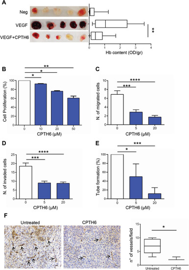
a Representative images of matrigel plugs and hemoglobin content of in vivo vessel formation assessed after injection of C57BL/6 mice with matrigel plugs containing PBS (Negative control), VEGF alone (Positive control) or VEGF in combination with 50 μM CPTH6. Five matrigel plugs/group of a representative experiment are shown. The values were expressed as optical density (OD)/gr matrigel plug. p-values were calculated between positive and CPTH6-containing matrigel plugs. b Analysis of proliferation of HUVEC exposed to increasing concentrations of CPTH6 for 72 h. The results are reported as percentage of “proliferation of CPTH6-treated cells/proliferation of control cells”. The results represent the average ± SD of three independent experiments. c,d Quantification of in vitro cell migration (c) and cell invasion (d) of HUVEC exposed to the indicated concentrations of CPTH6 for 6 h. The results are reported as average ± SD of number of migrated or invaded cells. e Quantification of capillary-like structure formation in HUVEC exposed to the indicated concentrations of CPTH6 for 6 h. The results are reported as percentage of tube formation relative to control, and represent the average ± SD of three independent experiments. f Representative images and relative quantification of immunohistochemical detection of microvessel density by CD31 staining in LCSC136 tumors from control or CPTH6-treated mice. NOD/SCID mice inoculated subcutaneously with 2.5 × 10⁵ LCSC136 cells and treated intraperitoneally with CPTH6 (50 mg/Kg; 5 days every 24 h for 3 weeks), IHC analysis was performed at the end of treatment. Magnification 20X. Scale bar, 30 μm. b-fp-values were calculated between control and treated cells. *p < 0.05; **p < 0.01; ***p < 0.001; ****p < 0.0001.
|
