|
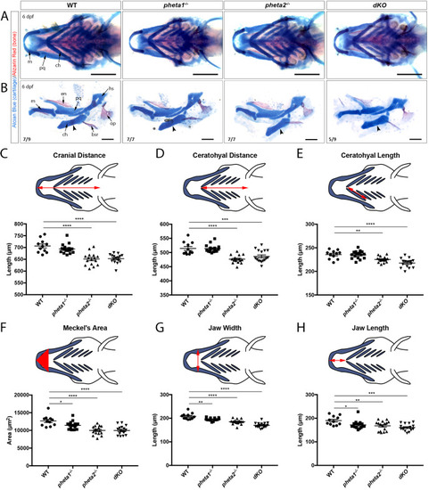
Loss of pheta1/2 disrupts craniofacial development. (A,B) Alcian Blue and Alizarin Red staining in 6 dpf animals. Structures are as indicated in the leftmost images. (A) Ventral view of the lower jaw. Representative images from each genotype are shown. Images are quantified in panels C-H. Scale bars: 200 µm. (B) Flat-mount preparations of the lower jaw at 6 dpf. Arrowheads point to where osteogenesis occurs within the ceratohyal cartilage. The number of animals imaged with displayed phenotype is shown in the lower-left corner of each image. Scale bars: 100 µm. (C-H) Morphological measurements of 6 dpf larvae, with the measured distance/area highlighted in red in the above schematics. bsr, brachiostegal ray; ch, ceratohyal; en, entopterygoids; hs, hyosymplectic; m, Meckel's cartilage; op, opercle; pq, palatoquadrate. *P<0.05, **P<0.01, ***P<0.001, ****P<0.0001.
|