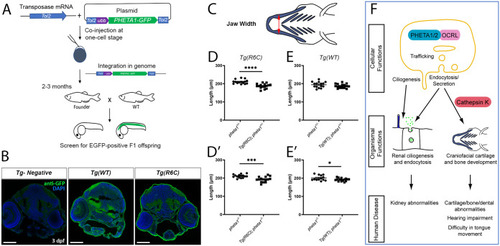
Pheta1R6C exerts a dominant-negative effect on craniofacial development in the partial or complete absence of Pheta1. (A) Outline of the procedures for generating the Tg(R6C) and Tg(WT) transgenic lines. (B) Confocal images showing broad expression of Pheta1WT-GFP and Pheta1R6C-GFP larvae in transverse cryosections, stained with anti-GFP (green) and DAPI (blue). A control larva with no transgene expression (Tg-Negative) is shown for comparison. Scale bars: 100 μm. (C-E′) Craniofacial measurements of 6 dpf larvae, with schematics shown in C. (D,E) Jaw width of pheta1+/– animals with and without Tg(R6C) (D) and Tg(WT) (E) transgenes. (D′,E′) Jaw width of pheta1−/− animals with and without Tg(R6C) (D′) and Tg(WT) (E′) transgenes. *P<0.05, ***P<0.001, ****P<0.0001. (F) Summary model. At the subcellular level, PHETA1/2 is known to interact with OCRL to regulate intracellular trafficking, ciliogenesis, endocytosis and secretion. These cellular functions likely enable PHETA1/2 to facilitate renal and craniofacial development, with the latter further requiring cathepsin K regulation. In humans, a deficiency in PHETA1 function potentially leads to abnormal development of the kidney and craniofacial structures. Other functional impairments such as hearing and tongue movement may also be associated with abnormal cartilage or bone formation.
|
