
Figure 3—figure supplement 1.
- ID
- ZDB-FIG-200530-26
- Publication
- Lee et al., 2020 - Tgfb3 collaborates with PP2A and Notch signaling pathways to inhibit retina regeneration
- Other Figures
- All Figure Page
- Back to All Figure Page
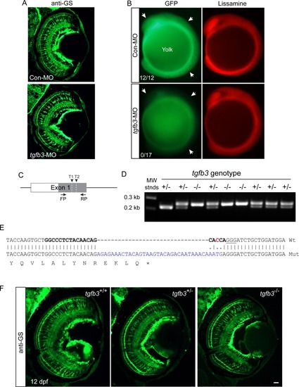
Tgfb3 knockdown and tgfb3 gene editing do not affect MG differentiation.(A) Effect of Tgfb3 knockdown on expression of MG differentiation at 6 dpf. Single cell zebrafish embryos were injected with the indicated MOs and assayed 6 days later for glutamine sythetase (GS) expression using immunofluorescence. (B) Representative images showing the consequences of experimental or control lissamine-tagged MO on reporter Tgfb3-EGFP expression at the 10 somite stage. Single cell embryos were injected with control or experimental MO along with tgfb3-EGFP RNA. At the 10 somite stage embryos were assayed for Tgfb3-EGFP expression using fluorescence microscopy. Arrows point to Tgfb3-EGFP expression in developing embryos that received control MO and to the reduced Tgfb3-EGFP expression in embryos that received experimental tgfb3-targeting MO. Numbers in panels indicate the number of lissamine+ embryos that also exhibited strong GFP expression. (C) Diagram of tgfb3exon one with position of gRNA1 and gRNA2 target sequences (T1 and T2) and primers used for PCR amplification across the mutation site. (D) Genotyping of interbred tgfb3+/- fish at 5 dpf. (E) DNA sequencing identifies an insertion/frame-shift mutation in tgfb3-/- fish at the gRNA2 target site. Shown is Wt and tgfb3 mutant (Mut) exon one sequence spanning gRNA2 target sequence (bold); predicted Cas9 cleavage site is indicated by a red C residue; pam sequence is underlined; blue sequence is insertion mutation; and asterisk indicates stop codon. (F) GS immunofluorescence in tgfb3+/+, tgfb3+/- and tgfb3-/- fish at 12 dpf shows normal MG differentiation in tgfb3+/- and tgfb3-/- fish. Shown is representative images of retinal sections with GS immunofluorescence. Size marker is 50 microns. |