
FIGURE
Fig. 4
- ID
- ZDB-FIG-200520-31
- Publication
- Kara et al., 2019 - The miR-216a-Dot1l Regulatory Axis Is Necessary and Sufficient for Müller Glia Reprogramming during Retina Regeneration
- Other Figures
- All Figure Page
- Back to All Figure Page
Fig. 4
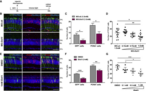
Dot1l Is Required for MG Dedifferentiation and Proliferation during Retina Regeneration (A) Control MO or dot1lMOs were injected and electroporated into the left eyes of Tg(1016tuba1a:gfp) zebrafish before intense light exposure (0 h). After 45 h, retinas were collected, sectioned, and immunostained using antibodies against GFP and PCNA. Nuclei were counterstained with TOPRO (blue). (B) Dot1l loss-of-function reduced tuba1a:GFP transgene expression and the number of INL PCNA+proliferating cells. (C) Quantification of GFP+dedifferentiated MG and PCNA+ proliferating progenitors in MO-ctl and MO-dot1l electroporated retinas. Data represent means ± SEMs, n = 5–6 fish; ∗∗p < 0.01 by 2-tailed Mann-Whitney U test. (D) Dose response to MO-dot1l. Increasing amounts of MO-dot1l MOs were injected and analyzed as in (B) and (C). Each data point represents an individual fish; ∗p < 0.05, ∗∗p < 0.01, and ∗∗∗p < 0.001 by 1-way ANOVA. (E) Control vehicle (DMSO) or iDot1l (Dot1l inhibitor EPZ004777) were injected intravitreally into the left eyes of Tg(1016tuba1a:gfp) zebrafish before intense light exposure (0 h). After 45 h, retinas were collected, sectioned, and immunostained using antibodies against GFP and PCNA. Nuclei were counterstained with TOPRO (blue). (F) Quantification of total GFP+ and PCNA+ cells. Data represent means ± SEMs, n = 10 fish; ∗p = 0.0111; ∗∗∗∗p < 0.0001 by 2-tailed Mann-Whitney Utest. (G) Dose response to iDot1l. Increasing amounts of the Dot1l inhibitor were injected and analyzed as in (E) and (F). Each data point represents an individual fish; ∗p < 0.05, ∗∗p < 0.01, and ∗∗∗p < 0.001 by 1-way ANOVA. GCL, ganglion cell layer; INL, inner nuclear layer; ONL, outer nuclear layer;. Scale bars, 50 μm. See also Figures S2 and S3. |
Expression Data
Expression Detail
Antibody Labeling
Phenotype Data
Phenotype Detail
Acknowledgments
This image is the copyrighted work of the attributed author or publisher, and
ZFIN has permission only to display this image to its users.
Additional permissions should be obtained from the applicable author or publisher of the image.
Full text @ Cell Rep.