
Fig. 10
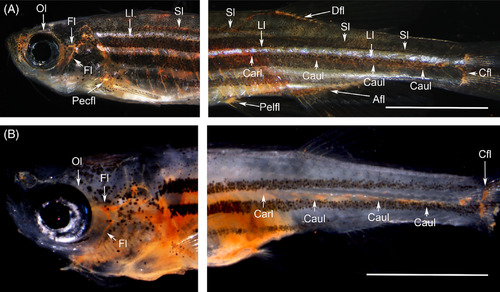
Functional lymph vascular system in the flt4 C526Δ (officially named flt4 um159) adult zebrafish mutant visualized through intraperitoneal dye injection (Abbreviations; see Table 1). A, Although subcutaneous color pigments interfered with visualization, all the functional lymph ducts were labeled with cinnabar in the wild‐type zebrafish (adult). B, Cinnabar dye labeled the branchial, facial and orbital lymph ducts in the cranial region but only weakly labeled the cardinal (thoracic duct) and caudal lymph vessels in the trunk and tail of the flt4 um159 mutant (3 months postfertilization). To verify each parietal lymphatic duct, the brain, gills, and visceral organs were removed from the wild‐type fish (A), but the gills and visceral organs remained in the mutant fish (B). Both fixed specimens were cleared with pure glycerol. White bars; 5 mm |