
Fig. 9
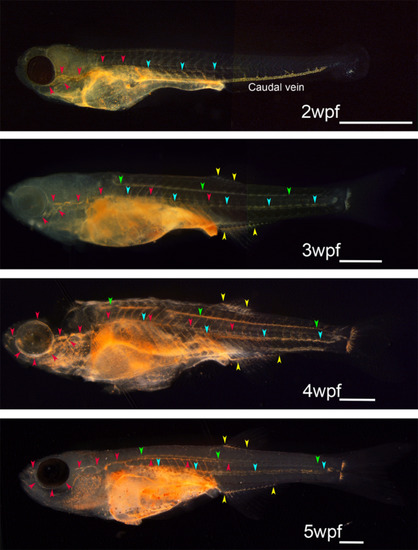
Developmental atlas of the functioning lymphatic vascular system in zebrafish obtained using intraperitoneal dye injection. Every functional lymphatic duct exhibits lymph flow and the ability to take up foreign particles in zebrafish. The functioning lymphatic vascular system was visualized by intraperitoneal dye injection in zebrafish at 2 to 5 wpf. Red arrowheads indicate superficial lymphatic ducts in the cranial region (facial and orbital lymphatics) and in the trunk (lateral lymphatic). Blue and green arrowheads indicate deep lymphatic ducts (cardinal‐caudal and spinal lymphatic, respectively). Yellow arrowheads indicate anal and dorsal fin lymphatics. Primitive caudal vein took up cinnabar particles actively during early developmental stages as shown in 2wpf zebrafish, but the phagocytotic character of the caudal vein was lost as embryonic lymphatic vascular system become functional. White bars; 1 mm |