FIGURE

Fig. S2

ID
ZDB-FIG-200513-19
Publication
Liu et al., 2020 - Eyes shut homolog (EYS) interacts with matriglycan of O-mannosyl glycans whose deficiency results in EYS mislocalization and degeneration of photoreceptors
Other Figures
All Figure Page
Back to All Figure Page
Fig. S2

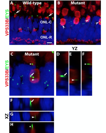
Mislocalized EYS in pomgnt1 mutant photoreceptors was not localized with late endosomes/lysosomes.
Cryosections of zebrafish eyes were double stained with anti-EYS (green) and with antibody against late endosome/lysosome marker VPS33B (red). The sections were counter-stained with DAPI to visualize nuclei (blue).

  1. (A)  Wild-type. In the wild-type retina, VPS33B antibody labeled the ellipsoid region of photoreceptors as well as apical portions of the outer plexiform layer. Most EYS immunoreactive puncta were observed on the apical side of VPS33B reactivity (arrows).

  2. (B)  Homozygous pomgnt1sny7 mutant. Most EYS immunoreactive puncta were localized in the outer nuclear layer. Some appeared to be over the VPS33B immunoreactive domain (arrowheads)

(C-H) Mutant. Max projection image and its orthogonal views of EYS puncta that appeared to be over the ATPB immunoreactive domains. Of 62 EYS immunoreactive puncta that appeared overlapping with VPS33B immunoreactivity, none were within the VPS33B immunoreactive domain, indicating that mislocalized EYS puncta were not located in late endosomes/lysosomes.
Scale bar in A: 4.36 
m for A-B, 2 m for C.

Expression Data

Expression Detail
Antibody Labeling
Phenotype Data

Phenotype Detail
Acknowledgments
This image is the copyrighted work of the attributed author or publisher, and ZFIN has permission only to display this image to its users. Additional permissions should be obtained from the applicable author or publisher of the image. Full text @ Sci. Rep.