
Fig. 5
- ID
- ZDB-FIG-200414-5
- Publication
- Ward et al., 2019 - Pharmacological restoration of visual function in a zebrafish model of von-Hippel Lindau disease
- Other Figures
- All Figure Page
- Back to All Figure Page
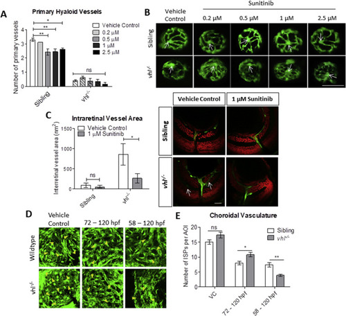
Sunitinib malate improves hyaloid vessel patterning, reduces retinal neovascularisation and inhibits excessive development of the choriocapillaris in vhl−/− larvae. A) Treatment from 58 hpf does not significantly increase vhl−/− primary hyaloid vessel number at 5 dpf. B) Representative images highlighting an overall improvement of vessel patterning following treatment with sunitinib from 58 hpf. White arrows denote central point from which primary blood vessels are counted. n = 3, 12 technical replicates per group per experiment. C) Reduction of ectopic intraretinal vasculature following treatment with 1 μM sunitinib. White arrows highlighting ectopic intraretinal vessels. Quantification of n = 5 larvae per treatment group. Scale bar = 20 μm. D) Z-stack projections of the sub-retinal choriocapillaris vasculature (endothelial cells shown in green) of Tg(fli1:EGFP) or vhl−/−(fli1:EGFP) at 120 hpf following treatment for 72 or 48 h as indicated. Yellow arrowheads point to interstitial pillars (ISPs). E) Quantification of the number of interstitial pillars per area of interest (AOI). n = 10 larvae. |
| Fish: | |
|---|---|
| Condition: | |
| Observed In: | |
| Stage: | Day 5 |
Reprinted from Developmental Biology, 457(2), Ward, R., Ali, Z., Slater, K., Reynolds, A.L., Jensen, L.D., Kennedy, B.N., Pharmacological restoration of visual function in a zebrafish model of von-Hippel Lindau disease, 226-234, Copyright (2019) with permission from Elsevier. Full text @ Dev. Biol.