
Figure 2-S1
- ID
- ZDB-FIG-200406-252
- Publication
- Xie et al., 2020 - Chemoptogenetic ablation of neuronal mitochondria in vivo with spatiotemporal precision and controllable severity
- Other Figures
- All Figure Page
- Back to All Figure Page
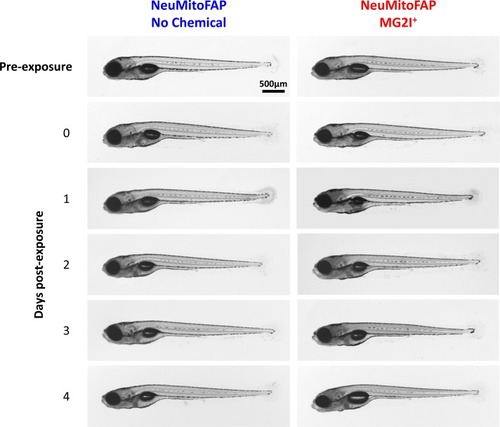
Transmitted light micrographs of NeuMitoFAP zebrafish: tThe right column shows larvae that were exposed to MG2I from 3 dpf; the left column shows untreated zebrafish. The first row shows larvae at 5dpf prior to light exposure. The second row shows larvae at 5dpf immediately after exposure to 60 J/cm2 light at λpeak=661nm. The subsequent rows show larvae at daily intervals (6–9 dpf) following light exposure at 5dpf. Although the MG2I-treated, light-exposed larvae showed loss of motor responses that did not recover during the time course of the experiment (see text), there were no major morphological abnormalities at any time point. |