|
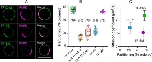
Relationship between diffusion in cells and analogue partitioning in phase-separated GPMVs.A, confocal images of TF-Chol, TF-PE, and TF-SM partitioning in phase separated GPMVs with rhodamine-PE (RhPE) as disordered phase marker. Scale bar is 10 μm. B, quantification of ordered domain partitioning of fluorescent lipid analogues from the confocal images. C, correlation between ordered domain partitioning in GPMVs and diffusion of fluorescent lipid analogues in cells. Data are shown as box-and-whisker plot showing median, first and third quartiles, and all the data values. Number of data points are indicated on the graphs.
|