
Fig. 1
- ID
- ZDB-FIG-200320-15
- Publication
- Prince et al., 2019 - Dorsal convergence of gastrula cells requires a Vangl2 and adhesion protein-dependent change in protrusive activity
- Other Figures
- All Figure Page
- Back to All Figure Page
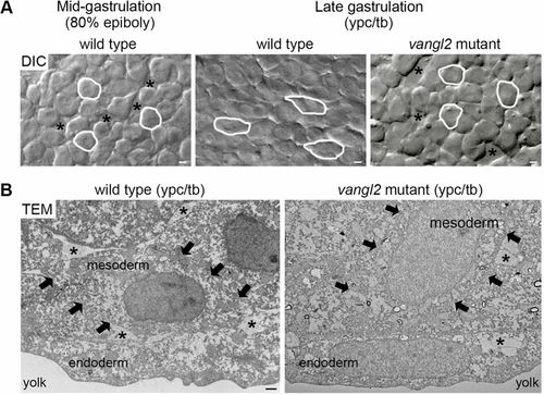
Vangl2-dependent changes in mesodermal cell behavior at late gastrulation. (A) Single-frame DIC time-lapse images highlighting morphological differences between wild-type mesendodermal cells at mid-gastrulation and mesodermal cells at late gastrulation (Jessen et al., 2002; Roszko et al., 2015). The mesoderm of a late gastrulation stage vangl2vu67/vu67 mutant embryo is shown for comparison. Images are oriented as shown in Fig. 2A with dorsal to the right and anterior to the top. Selected cells are outlined to show elongation and alignment relative to the dorsal-ventral body axis. Asterisks mark intercellular gaps. Scale bars: 5 µm. (B) Normal and defective PCP, as viewed in cross-sectioned images (6500× magnification) of late gastrulation wild-type and vangl2m209/m209 mutant deep mesodermal cells taken using transmission electron microscopy (TEM). Black arrows indicate the boundaries of single mesodermal cells. Asterisks indicate the presence of ECM between cells. Scale bar: 1 µm. |