
Fig. 5
- ID
- ZDB-FIG-200320-12
- Publication
- Daniel et al., 2019 - Spatiotemporal expression profile of embryonic and adult ankyrin repeat and EF-hand domain containing protein 1-encoding genes ankef1a and ankef1b in zebrafish
- Other Figures
- All Figure Page
- Back to All Figure Page
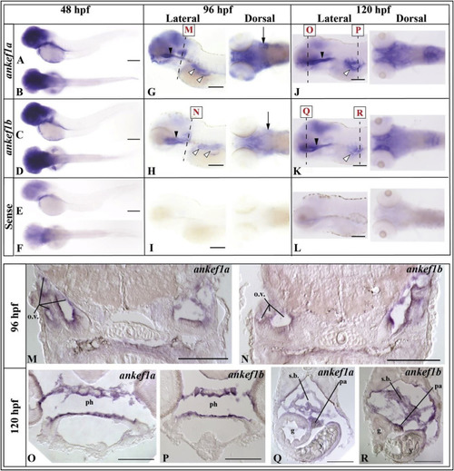
ankef1a and ankef1b expression in 48, 96, and 120 hpf embryos. A,C,E are lateral views, while B,D, and F are dorsal views. Black arrowheads in G-H and J-K indicate pharyngeal staining. Staining in the swim bladder region in 96 and 120 hpf is denoted by white arrowheads in G-H and J-K. Arrows in G-H point to otic vesicles. Scale bars for each whole mount images A-L = 200 μm. Relative whole-mount positions corresponding to cryosections M-N are indicated by dashed line and adjacent boxed red letters in G-H and J-K. M-N demonstrate ankef1a and ankef1b expression in the otic vesicles of a 96 hpf embryos. O–P show ankef1a and ankef1b expression in the pharynx of 120 hpf embryos. Q-R indicate ankef1a and ankef1b expression in the swim bladder of 120 hpf embryos. g: gut; o.v.: otic vesicle; pa: pancreas; ph: pharynx; s.b.: swim bladder; y: yolk. Scale bars for cryosections M-R = 100 μm. |
| Genes: | |
|---|---|
| Fish: | |
| Anatomical Terms: | |
| Stage Range: | Long-pec to Day 5 |
Reprinted from Gene expression patterns : GEP, 34, Daniel, J.G., Panizzi, J.R., Spatiotemporal expression profile of embryonic and adult ankyrin repeat and EF-hand domain containing protein 1-encoding genes ankef1a and ankef1b in zebrafish, 119069, Copyright (2019) with permission from Elsevier. Full text @ Gene Expr. Patterns