
Figure 1
- ID
- ZDB-FIG-200318-10
- Publication
- Weinberger et al., 2020 - Functional Heterogeneity within the Developing Zebrafish Epicardium
- Other Figures
- All Figure Page
- Back to All Figure Page
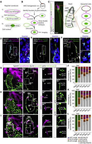
Heterogeneous Expression of (A) Fluorescence of (B) Workflow to establish (D–F) Single optical sections of mRNA stainings of (D) (G–I) Projections of the heart in triple reporter larvae at 3 dpf (G), 5 dpf (H), and 7 dpf (I). (G′ and H′) Single optical sections from (G) and (H). (I′) Short projection from (I). (G″–I″ and G‴–I‴) Representative epicardial fluorescence patterns. (J–L) Relative quantification of epicardial fluorescence patterns across the regions indicated in (C) at 3 dpf (J), 5 dpf (K), and 7 dpf (L). Scale bars: 20 μm in (D)–(F), 5 μm in (D′)–(F′), 50 μm in (G)–(I) and (G′)–(I′), and 10 μm in (G″)–(I″) and (G‴)–(I‴). Color channels were adjusted separately for brightness and contrast. Data in (J)–(L) are represented as mean minus standard deviation. Number of embryos analyzed: 3 dpf n = 6, 5 dpf n = 10, and 7 dpf n = 6. V, ventricle; BA, bulbus arteriosus. See also |
| Genes: | |
|---|---|
| Fish: | |
| Anatomical Terms: | |
| Stage Range: | Protruding-mouth to Days 7-13 |
Reprinted from Developmental Cell, 52(5), Weinberger, M., Simões, F.C., Patient, R., Sauka-Spengler, T., Riley, P.R., Functional Heterogeneity within the Developing Zebrafish Epicardium, 574-590.e6, Copyright (2020) with permission from Elsevier. Full text @ Dev. Cell