
Fig. 7
- ID
- ZDB-FIG-200311-28
- Publication
- Zhu et al., 2019 - Migratory Neural Crest Cells Phagocytose Dead Cells in the Developing Nervous System
- Other Figures
- All Figure Page
- Back to All Figure Page
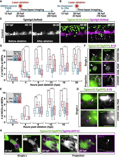
NCC Recruitment toward Damage Is Mediated by Il-1β Signaling (A) Schematic diagram of experimental design in (D), which is YVAD treatment versus control. (B) Schematic diagram of experimental design in (E), which is IL-1Ra treatment versus control. (C) Images from a time-lapse movie of a Tg(sox10:nls-Eos);Tg(olig2:DsRed) embryo before and after ablation of two motor neurons (asterisks). Dashed box indicates the region where the numbers of NCCs are counted in (D) and (E). (D) Quantification of the number of NCCs in DMSO or YVAD-treated embryos after ablation. Numbers in the legends denote the number of embryos quantified. ****p ≤ 0.0001; ***p ≤ 0.001; **p ≤ 0.01; *p ≤ 0.05. Same for (E). (E) Quantification of the number of NCCs in control embryos or embryos treated with IL-1Ra after motor neuron ablation. (F) Images from 20 hpf Tg(sox10:TagRFP) embryos labeled with an Il-1β antibody; NCC protrusions (arrows), NCC debris (filled arrowheads), and NCC engulfment vesicles (open arrowheads). (G) In 20 hpf Tg(sox10:TagRFP) embryos labeled with an Il-1β antibody, NCC vesicles are filled with Il-1β+ debris (arrowheads). (H) Images from Tg(sox10:TagRFP);il1b:GFP-F embryos at 20 hpf. Open arrowheads denote phagocytic NCCs that are il-1β+. Filled arrowheads denote il-1β+ debris inside a NCC vesicle. Scale bars, 20 μm in (C), 10 μm in (F)–(H). |
Reprinted from Cell, 179(1), Zhu, Y., Crowley, S.C., Latimer, A.J., Lewis, G.M., Nash, R., Kucenas, S., Migratory Neural Crest Cells Phagocytose Dead Cells in the Developing Nervous System, 74-89.e10, Copyright (2019) with permission from Elsevier. Full text @ Cell