FIGURE

Fig. 4

ID
ZDB-FIG-200306-68
Publication
Truong et al., 2020 - High-contrast, synchronous volumetric imaging with selective volume illumination microscopy
Other Figures
All Figure Page
Back to All Figure Page
Fig. 4

Functional neuroimaging with SVIM.

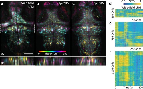
Functional imaging of a 5 dpf larval zebrafish with pan-neuronal fluorescent calcium indicators, Tg(elavl3:H2b-GCaMP6s). Spontaneous brain activity, over a volume ~600 × 600 × 100 µm3 (depth), was recorded at 1 volume s−1, with SVIM, in either 1- or 2-photon excitation mode (1p-SVIM or 2p-SVIM, respectively), or conventional 1p wide-field LFM. Cellular-resolution representations of active neurons were found with standard methodology based on spot segmentation of the time-domain standard deviation of the 3D time-series data (Methods section). ac Images shown are depth color-coded xy- or xz-projections, of the time-domain standard deviation projection of the recorded brain activity over a time window of 100 s. Colored ellipsoids represent active neurons. Dashed box in the xy-projection image represent the region that produces the corresponding xz-projection image. Activity traces of segmented neurons are shown in df, revealing that the most number of neurons were found with 2p-SVIM (1104 cells), then with 1p-SVIM (796 cells), both of which were several-fold higher than with conventional wide-field LFM (263 cells). Scale bars, 100 µm.

Expression Data

Expression Detail
Antibody Labeling
Phenotype Data

Phenotype Detail
Acknowledgments
This image is the copyrighted work of the attributed author or publisher, and ZFIN has permission only to display this image to its users. Additional permissions should be obtained from the applicable author or publisher of the image. Full text @ Commun Biol