|
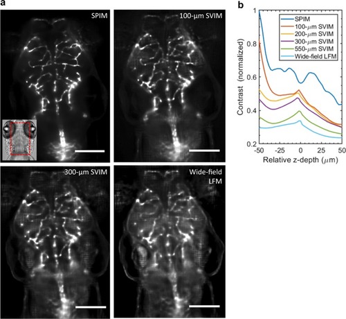
Higher contrast achieved by SVIM.a SVIM images of the cranial vasculature improve in contrast as the depth (axial extent) of the illumination volume is decreased. Images are averaged-intensity z-projections of the same 40-µm thick sub-volume, centered at ~170 µm into the head of a 5 dpf zebrafish larva. The SVIM image quality progressively approaches the performance of SPIM as the axial extent of the illumination is reduced to 300 µm or 100 µm, far exceeding the image contrast obtained with wide-field LFM. Inset shows the approximate location of the imaged volume, in context of the zebrafish head. b Quantitative comparison of image contrast, defined as the normalized standard deviation of the pixel values (Methods section), comparing LFM, SPIM, and SVIM of different SVI extents from a. SVIM of smaller extents yielded increasingly better contrast, approaching the performance of SPIM. The contrast of SPIM showed the intrinsic contrast variation of the 3D sample, coupled with the expected contrast decay for increasing imaging depth. The local increase in contrast seen for the SVIM and LFM cases around z = 0 µm came from grid-like artifacts from the light-field reconstruction centered around the native focal plane, a known feature of LFM in general3,4. Scale bars, 100 µm.
|