
Fig. 6
- ID
- ZDB-FIG-200207-17
- Publication
- Mourabit et al., 2019 - New insights into organ-specific oxidative stress mechanisms using a novel biosensor zebrafish
- Other Figures
- All Figure Page
- Back to All Figure Page
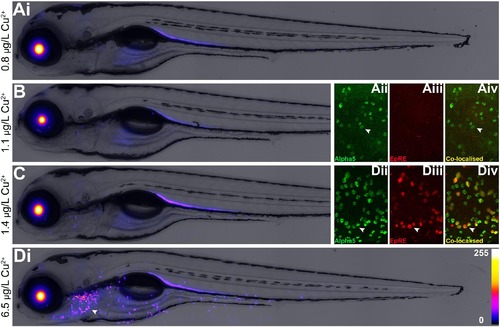
Concentration-dependent tissue response in Tg (3EpRE:hsp70:mCherry) - zebrafish larvae exposed to Cu2+. Fish larvae were exposed to either a control (0.83 μg/L Cu2+; Ai-iv) or three concentrations of Cu2+ (B; C; Di-iv) from 2 to 4 dpf and subsequently imaged via epifluorescence and confocal microscopy. Confocal microscopic images of fixed larvae under control (Aii-iv) and 6.46 μg/L Cu2+ (Dii-iv) conditions; green fluorescence indicates staining with an anti-Na+ K+ ATPase antibody (alpha5), red fluorescence is EpRE-mCherry signal, and yellow indicates co-localised signal. Arrowheads = ionocytes. For images (A–D), fluorescence intensity is represented using a “Fire” lookup table with a 0–255 pixel intensity calibration bar. Measured Cu2+ culture medium concentrations displayed. (For interpretation of the references to color in this figure legend, the reader is referred to the web version of this article.) |