
Fig. 2
- ID
- ZDB-FIG-200207-13
- Publication
- Mourabit et al., 2019 - New insights into organ-specific oxidative stress mechanisms using a novel biosensor zebrafish
- Other Figures
- All Figure Page
- Back to All Figure Page
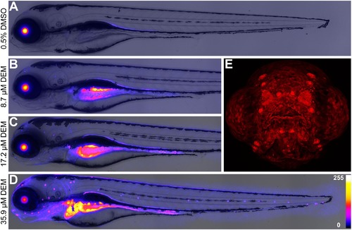
Concentration-dependent tissue fluorescence in Tg (3EpRE:hsp70:mCherry) zebrafish larvae exposed to diethylmaleate (DEM). Fish larvae were exposed to solvent control (A) and three concentrations of DEM (B; C; D) from 2 to 4 dpf, and subsequently imaged via epifluorescence and confocal microscopy. A confocal image of the frontal head region (E) is presented to illustrate the range of surface tissues (e.g. skin cells, neuromasts, olfactory pits) affected by exposure to 35.9 μM DEM. For images (A–D), fluorescence intensity is represented using a “Fire” lookup table with a 0–255 pixel intensity calibration bar. Measured DEM E3 culture medium concentrations displayed. |