
Fig. 1
- ID
- ZDB-FIG-200130-41
- Publication
- Dasyani et al., 2019 - Lineage tracing of col10a1 cells identifies distinct progenitor populations for osteoblasts and joint cells in the regenerating fin of medaka (Oryzias latipes)
- Other Figures
- All Figure Page
- Back to All Figure Page
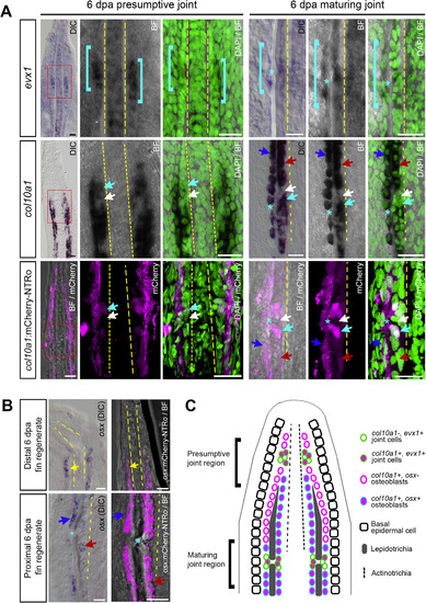
Expression of evx1, col10a1, osx, col10a1:mCherry-NTRo and osx:mCherry-NTRo in the adult medaka fin. A: Comparison of endogenous evx1 and col10a1 expression by RNA in situ hybridization and transgenic col10a1:mCherry-NTRo expression in the presumptive joint region and maturing joint region of 6 dpa fin regenerates (20 μm longitudinal sections). Endogenous col10a1 expression and col10a1:mCherry-NTRo expression mark a subset of evx1-positive joint cells (cyan arrows, n = 3 fin rays, 3 fish). Cyan brackets indicate domains of evx1-positive joint cells, asterisks mark joints, yellow dotted lines mark the actinotrichia, cyan arrows mark col10a1 expression in joint cells, white arrows mark absence of expression in joint cells, dark blue arrows mark the outer row of osteoblasts, red arrows mark the inner row of osteoblasts. B: Comparison of endogenous osx expression by RNA in situ hybridization and transgenic osx:mCherry-NTRo expression in distal and proximal regions of 6 dpa fin regenerates (20 μm longitudinal sections). osx:mCherry-NTRo expression recapitulates endogenous osx expression, marking a single row of osteoblasts (yellow arrows) per hemiray at the distal fin regenerate and two rows of osteoblasts (dark blue and red arrows) per hemiray in the proximal regenerate (n = 3 fin rays, 3 fish). Scale bars = 20 μm. C: Schematic diagram indicating expression patterns of evx1, col10a1, and osx in joint cells and osteoblasts at the distal region of a 6 dpa fin regenerate in a longitudinal section view. |
Reprinted from Developmental Biology, 455(1), Dasyani, M., Tan, W.H., Sundaram, S., Imangali, N., Centanin, L., Wittbrodt, J., Winkler, C., Lineage tracing of col10a1 cells identifies distinct progenitor populations for osteoblasts and joint cells in the regenerating fin of medaka (Oryzias latipes), 85-99, Copyright (2019) with permission from Elsevier. Full text @ Dev. Biol.