|
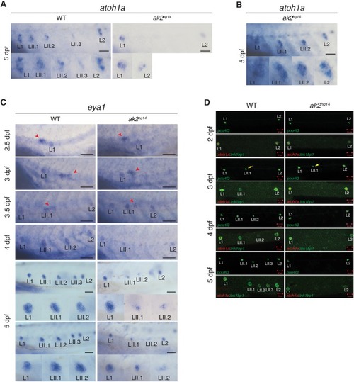
Characterization of PLL neuromast markers in lateral line of ak2hg14 mutants. (A,B) WISH analysis of atoh1a marker expression in primary and secondary neuromasts on 5 dpf ak2hg14 (A) and ak2hg16 (B) embryos and their siblings (WT). (C) eya1 expression from 2.5 to 5 dpf in ak2hg14 embryos and their siblings. Red arrowheads indicate the primII. Each panel shows higher magnification of the regions in dashed rectangles from corresponding pictures in Fig. S5. Scale bars: 100 µm. (D) Confocal analysis from 2 to 5 dpf of trunk regions of ak2hg14 embryos and their siblings with different transgenic backgrounds to visualize components of the lateral line rosettes. Scale bars: 50 µm. Yellow arrows indicate pou4f3:GFP+ HCs in secondary neuromasts.
|