|
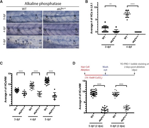
Reduced hair cell number and secondary neuromast differentiation phenotypes in ak2hg14 mutants. (A) Alkaline phosphatase staining of 3-5 dpf ak2hg14 homozygous embryos and their siblings (WT). White arrowheads indicate the migrating secondary primordium (primII). L1-2 and LII.1-3 designate primary and secondary neuromasts, respectively. Scale bars: 50 μm. (B,C) Yo-PRO-1 iodide staining of hair cells (HCs) in lateral line neuromasts in ak2hg14 embryos and their wild-type siblings during development to compare the average number of HCs in LII.1 secondary neuromasts (B) and in primary neuromasts (C). (D) Average number of HCs per primary neuromast in Yo-PRO-1 iodide-stained ak2hg14 embryos and their siblings 2 days after ablation with CuSO4. Mean±s.d.**P<0.0011; ****P<0.0001 (unpaired, two-tailed Student's t-test).
|