FIGURE

Fig. 4

ID
ZDB-FIG-191230-93
Publication
Taylor et al., 2019 - Adaptive prospective optical gating enables day-long 3D time-lapse imaging of the beating embryonic zebrafish heart
Other Figures
All Figure Page
Back to All Figure Page
Fig. 4

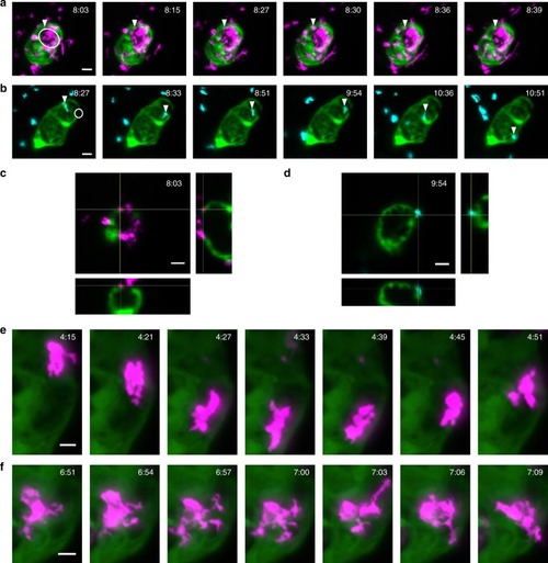
Sustained beating-heart time-lapse imaging of immune cell responses to cardiac injury. a, b Sequence of MIPs (from Supplementary Videos 68) showing macrophages (a magenta mpeg1:mCherry) and neutrophils (b cyan mpx:mCherry) migrating to, and interacting at, the laser wound site (white circle) on the ventricle (myl7:GFP, laser injury has bleached myocardial cells at the injury site). Arrowheads mark tracked immune cells interacting with the wound margins, changing shape and reverse migrating from the wound to the pericardium. c, d Orthogonal plane views co-localising the tracked macrophages (c) and neutrophils (d) on and within the myocardium at the wound site. e, f Individual macrophages are seen to undergo complex shape changes during migration. All timestamps show hours post injury (hpi). Scale bars: 30 μm (10 μm for e, f)

Expression Data

Expression Detail
Antibody Labeling
Phenotype Data

Phenotype Detail
Acknowledgments
This image is the copyrighted work of the attributed author or publisher, and ZFIN has permission only to display this image to its users. Additional permissions should be obtained from the applicable author or publisher of the image. Full text @ Nat. Commun.