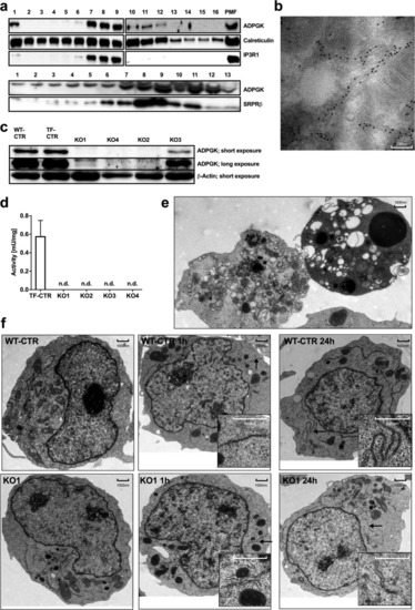
ADPGK is localized in ER lumen and important for ER biogenesis. (a) Representative immunoblots of density gradient-enriched ER fractions from Jurkat T cells, stained for ADPGK and different ER-markers (IP3R-1, Inositol-1,4,5-triphosphate receptor; SRPRβ, signal recognition particle receptor subunit β; PMF, post-mitochondrial fraction). N = 4 independent experiments. (b) Representative electron micrograph of ADPGK-GFP expressing HEK cells, stained with gold particle-labeled anti-GFP antibodies. (c) Representative immunoblots of ADPGK protein in Jurkat T cell knockout using β-Actin as a loading control. N = 5 independent experiments. (d) ADPGK activity assays in KO clones normalized to protein content. N = 7 independent experiments. (e) Electron micrographs of KO1 cells stimulated with PMA (10 ng/ml) and Ionomycin (10 µM) for 24 h. Dying cells show features of autophagy (left) and apoptosis (right). (f) Electron micrographs of KO1 and WT-CTR cells stimulated with PMA (10 ng/ml) and Ionomycin (10 µM) for 0 h, 1 h, and 24 h. Stimulation results in extended ER networks in control cells and short, dilated ER structures in KO1 cells. Black arrows indicate magnified structures. All images of blots represent cropped blots of appropriate protein size. For full length blots see Supplemental Fig. 3.
|
