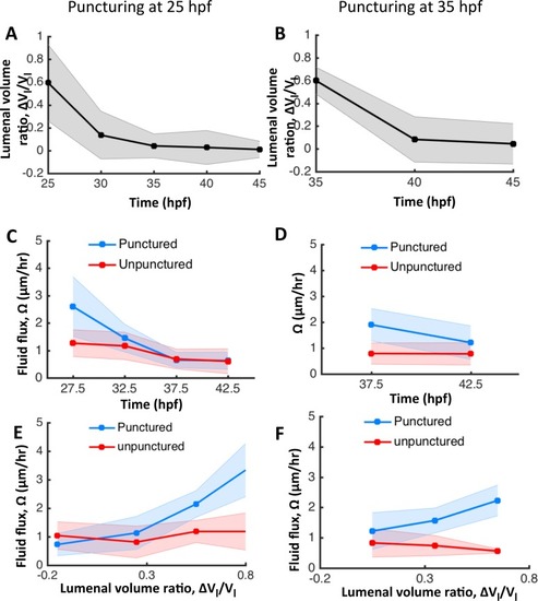
Figure 4—figure supplement 1.

The otic vesicle regenerates to stage-specific volumes when punctured between 25–45 hpf.

In addition to experiments at 30 hpf reported in Figure 4, puncturing was conducted at 25 hpf (A, C, E) and 35 hpf (B, D, F). (A–B) Volume regeneration and establishment of contralateral symmetry after unilateral puncturing. The y-axis plots the difference in lumenal volumes normalized to the unpunctured volume (ΔVlVl). (C–D) Lumenal fluid flux Ω in punctured (blue) and unpunctured (red) vesicles during regeneration showing the increase in Ω in punctured embryos and gradual decay to the unpunctured levels. (E–F) Plot showing Ω as a function of ΔVlVl. The data was pooled across multiple embryos for the punctured (blue) and unpunctured (red) ears throughout their regeneration period until 45 hpf. Ω in punctured ears (blue) is positively correlated with ΔVlVl while unpunctured ears (red) continued growing with an approximately constant flux.

Expression Data

Expression Detail
Antibody Labeling
Phenotype Data

Phenotype Detail
Acknowledgments
This image is the copyrighted work of the attributed author or publisher, and ZFIN has permission only to display this image to its users. Additional permissions should be obtained from the applicable author or publisher of the image. Full text @ Elife