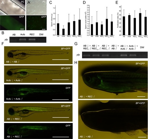
Production of donor-derived spermatozoa and progeny from MO-sterilized recipients. Milt (A) and individual spermatozoa (A’) stripped from recipients demonstrating yfp fluorescence. (B) RT-PCR amplification of yfp in milt obtained from wild-type AB (AB), β-actin transgenic (pku341Tg; Actb) and recipient (REC) zebrafish. Milt volume (C) and sperm count (D) of milt obtained from wild-type AB (AB), β-actin transgenic fish (pku341Tg; Actb) and recipients of fresh (RECF), frozen (RECSR) and vitrified (RECV) spermatogonia. (E) Fertilization rates after spawning control fish as well as recipient males and wild-type AB females. (F) Three dpf larvae obtained from crossing control fish and recipient males with wild-type AB females under fluorescent stereomicroscope. (G) PCR amplification of yfp in offspring obtained from control crossings, as well as from crossing recipient males with wild-type AB females. (H) F1 individuals developed normally and donor-derived individuals displayed fluorescent signal compared to the control crossing six months after fertilization. Values in C–E are presented as mean ± SD. Lack of different letters above SD lines indicate the lack of statistical significance (Tukey’s HSD, p > 0.05). Scale bars: (A) 100 µm; (A’) 20 µm; (F) 1 mm; (H) 2.5 mm.
|
