FIGURE

Figure 2

ID
ZDB-FIG-191230-245
Publication
Marinović et al., 2019 - Preservation of zebrafish genetic resources through testis cryopreservation and spermatogonia transplantation
Other Figures
All Figure Page
Back to All Figure Page
Figure 2

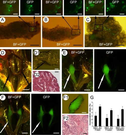
Incorporation and proliferation of fresh and cryopreserved spermatogonia. The incorporation and proliferation of fresh (A) and cryopreserved (B,C) vasa::egfp spermatogonia within the testes (A,B) and ovaries (C) of non-sterilized recipients. Testes (arrows) of the control MO-sterilized recipients (D) appear undeveloped under the stereomicroscope (D1), while the histological analysis (D2) displayed a lack of developing germ cells. Recipients of actb:yfp spermatogonia displayed either one (E) or both (F) developed testes (arrows). Developed testes displayed strong green fluorescence originating from donor actb:yfp spermatogonia (F1), while histological analyses displayed clear differentiation of germ cells into spermatozoa (F2). (G) Incorporation of fresh and cryopreserved spermatogonia into non-sterilized (NST) or MO-sterilized (ST) recipients. Values are presented as mean ± SD. Scale bars: (A, B, C) 500 µm; (top panels) 100 µm; (D, E, F) 1 mm; (D1, F1) 200 µm; (D2, F2) 20 µm.

Expression Data

Expression Detail
Antibody Labeling
Phenotype Data

Phenotype Detail
Acknowledgments
This image is the copyrighted work of the attributed author or publisher, and ZFIN has permission only to display this image to its users. Additional permissions should be obtained from the applicable author or publisher of the image. Full text @ Sci. Rep.