FIGURE

Figure 2—figure supplement 1

ID
ZDB-FIG-191230-1582
Publication
Morishita et al., 2019 - A critical role of VMP1 in lipoprotein secretion
Other Figures
All Figure Page
Back to All Figure Page
Figure 2—figure supplement 1

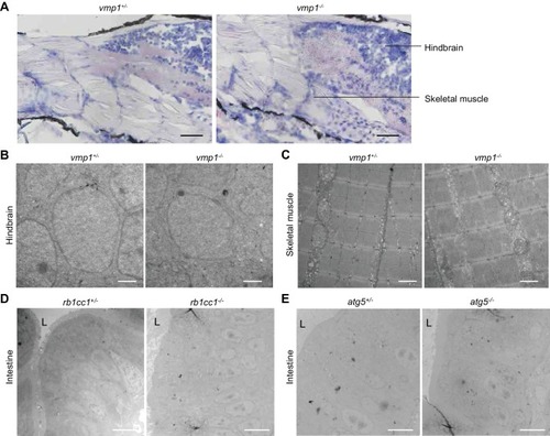
Large lipid-containing structures are not observed in the brain and skeletal muscle of vmp1-/- zebrafish or in the intestine of rb1cc1-/- and atg5-/- zebrafish.

(A) Oil red O and hematoxylin staining of 6-dpf vmp1+/- and vmp1-/- zebrafish. Data are representative of two independent experiments. Scale bars, 100 μm. (B) and (C) Transmission electron microscopy of the hindbrain (B) and skeletal muscle (C) from 6-dpf vmp1+/- and vmp1-/- zebrafish. Data are representative of two independent experiments. Scale bars, 1 μm. (D) and (E) Transmission electron microscopy of the intestine from 6-dpf rb1cc1+/-, rb1cc1-/- (D), atg5+/- and atg5-/- (E) zebrafish. L, lumen of the intestine. Data are representative of two independent experiments. Scale bars, 5 μm.

Expression Data

Expression Detail
Antibody Labeling
Phenotype Data
Fish:
Observed In:
Stage: Day 6

Phenotype Detail
Acknowledgments
This image is the copyrighted work of the attributed author or publisher, and ZFIN has permission only to display this image to its users. Additional permissions should be obtained from the applicable author or publisher of the image. Full text @ Elife