FIGURE

Fig 4

ID
ZDB-FIG-191230-1073
Publication
Marquart et al., 2019 - Prepontine non-giant neurons drive flexible escape behavior in zebrafish
Other Figures
All Figure Page
Back to All Figure Page
Fig 4

Prepontine escape neurons project reciprocally to the caudal hindbrain and cerebellum.

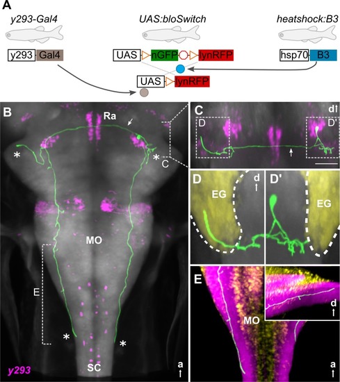
(A) Schematic of 3 transgene system used for B3-recombinase–based neuronal tracing. (B-E) Representative traced neuron in 6-dpf larva (green, for others, see S5 Fig), registered to ZBB atlas [18]. Background is elavl3:Cer (gray). (B) Horizontal maximum whole-brain projection of a reconstructed neuron from y293-Gal4 (ZBB, magenta). Asterisks: projections of the 4 primary neurites. Arrow: commissural projection. Dashed lines indicate views in (C) and (E). (C) Coronal substack projection from the area indicated in (B). Arrow: commissural projection. Scale bar 50 μm. Views in (D) outlined. (D) Coronal projections of neurites extending into the ipsilateral (D') and contralateral (D) EG (yellow). (E) Dorsal projection through the caudal medulla lateral neuropil area (ZBB anti-zrf2, purple). Cellular regions labeled by Tg(elavl3:nls-mCar)y517 (ZBB, yellow). Inset: sagittal view of same region. a, anterior; d, dorsal; EG, eminentia granularis; hsp70, heat-shock protein 70; lynRFP, membrane-tagged red fluorescent protein; MO, medulla oblongata; nGFP, nuclear-localized green fluorescent protein; Ra, raphe; SC, spinal cord; ZBB, Zebrafish Brain Browser.

Expression Data

Expression Detail
Antibody Labeling
Phenotype Data

Phenotype Detail
Acknowledgments
This image is the copyrighted work of the attributed author or publisher, and ZFIN has permission only to display this image to its users. Additional permissions should be obtained from the applicable author or publisher of the image. Full text @ PLoS Biol.