
Fig. 2
- ID
- ZDB-FIG-190823-21
- Publication
- Whitesell et al., 2019 - foxc1 is required for embryonic head vascular smooth muscle differentiation in zebrafish
- Other Figures
- All Figure Page
- Back to All Figure Page
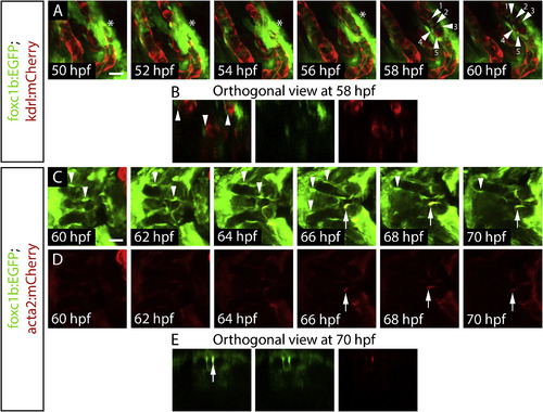
Timelapse of foxc1b expressing mesenchymal cells attaching to the endothelium followed by upregulation of acta2. A) Ventral views of aortic arch arteries of a foxc1b:EGFP; kdrl:mCherry embryo using confocal time-lapse imaging starting at 50 hpf. Mesenchymal cells are indicated by the asterisk, while vascular mural cells are indicated by arrowheads, starting by 56–58 hpf. B) Orthogonal views at 58 hpf depict the foxc1b:EGFP cells wrapping around, but not co-expressing an endothelial marker. C) Time-lapse of the early ventral aorta of a foxc1b:EGFP; acta2:mCherry embryo from 60 to 70 hpf. foxc1b:EGFP positive smooth muscle cells are associated with the endothelium (arrowheads) but do not co-express acta2:mCherry until approximately 66 hpf (arrow). D) Single channel images of acta2:mCherry. E) Orthogonal views at 70 hpf show acta2:mCherry co-expressed within a foxc1b:EGFP positive cell. Scale bars represent 20 μm. |
| Genes: | |
|---|---|
| Fish: | |
| Anatomical Terms: | |
| Stage Range: | Long-pec to Pec-fin |
Reprinted from Developmental Biology, 453(1), Whitesell, T.R., Chrystal, P.W., Ryu, J.R., Munsie, N., Grosse, A., French, C.R., Workentine, M.L., Li, R., Zhu, L.J., Waskiewicz, A., Lehmann, O.J., Lawson, N.D., Childs, S.J., foxc1 is required for embryonic head vascular smooth muscle differentiation in zebrafish, 34-47, Copyright (2019) with permission from Elsevier. Full text @ Dev. Biol.