
Fig. S2
- ID
- ZDB-FIG-190730-6
- Publication
- Andalman et al., 2019 - Neuronal Dynamics Regulating Brain and Behavioral State Transitions
- Other Figures
- All Figure Page
- Back to All Figure Page
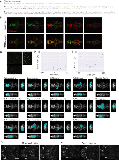
Registration and Resolution of LFM Volumes, Related to Figure 2 (A) The three Computation Morphometry Toolkit (CMTK) commands used to register LFM volumes to a common reference atlas. Input filenames for each command are displayed in blue text; output filenames are displayed in red text; and optional parameters are displayed in green text. (B) Five evenly spaced horizontal slices from a LFM volume of an experimental fish (green) and the LFM atlas (red) before (top) and after (bottom) non-rigid registration using CMTK. (C-E) Assessment of spatial resolution of LFM. (C) Orthogonal maximum intensity projections of a light field volumetric reconstruction of 1μm fluorescent beads embedded in 1% agarose (100 μm scale bar; xy projection in top left; 200 ms exposure time; 6.3 mW excitation light). (D and E) The lateral (D) and axial (E) full width at half maximum of individual fluorescent beads located at different depths within the volume displayed in panel C. These data provide an upper bound on the resolution of our LFM microscope because the Richarson-Lucy deconvolution algorithm is optimal suited for sparse samples such as a bead volume (Broxton et al., 2013, Shaked et al., 2011). For comparison, the theoretical diffraction-limited FWHM of the point spread function is 0.437 μm laterally and 3.176 μm axially when using 515 nm light and a 0.6 NA objective. (F) Atlas region masks (teal) are shown overlaid on orthogonal maximum intensity projections of the LFM atlas. (G and H) 2P stack of in situ hybridation for c-fos max-projected over 15 μm within the vHb in three fish 30 min after the end of BC with 5V shocks (G) and no shocks (H; 0V; blue triangle demarcate lateral edges of vHb; scale bar, 100 μm). |
Reprinted from Cell, 177(4), Andalman, A.S., Burns, V.M., Lovett-Barron, M., Broxton, M., Poole, B., Yang, S.J., Grosenick, L., Lerner, T.N., Chen, R., Benster, T., Mourrain, P., Levoy, M., Rajan, K., Deisseroth, K., Neuronal Dynamics Regulating Brain and Behavioral State Transitions, 970-985.e20, Copyright (2019) with permission from Elsevier. Full text @ Cell