
FIGURE
Fig. 5
- ID
- ZDB-FIG-190726-68
- Publication
- Colucci-Guyon et al., 2019 - Ultraspecific live imaging of the dynamics of zebrafish neutrophil granules by a histopermeable fluorogenic benzochalcone probe
- Other Figures
- All Figure Page
- Back to All Figure Page
Fig. 5
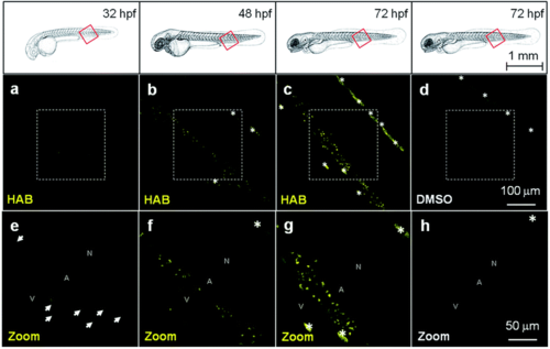
HAB labels specific cells in live zebrafish from 32 hpf. Confocal fluorescence imaging of HAB labeling (10 μM) in live wild-type zebrafish embryos (32 and 48 hpf) and swimming larvae (72 hpf) following excitation at 488 nm and detection in the 550–650 nm range. The yellow-orange color is indicative of the fluorescence seen with the naked eye. Maximum intensity Z-projection images (2 μm serial optical sections) are shown. Arrows point to the HAB label; asterisks mark pigment cells. A, artery; N, notochord; V, vein. |
Expression Data
Expression Detail
Antibody Labeling
Phenotype Data
Phenotype Detail
Acknowledgments
This image is the copyrighted work of the attributed author or publisher, and
ZFIN has permission only to display this image to its users.
Additional permissions should be obtained from the applicable author or publisher of the image.
Full text @ Chem Sci