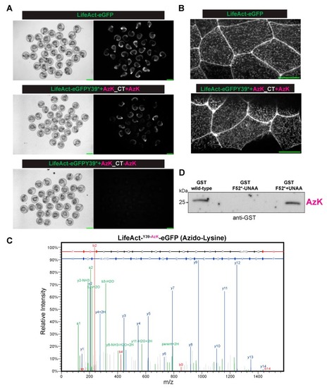
The site-specific incorporation of azido-lysine in the zebrafish embryos. (A) The Brightfield (left) or fluorescent (right) images of the zebrafish embryos injected with mRNA(s) for LifeAct-eGFP (top panel) or LifeAct-eGFP Y39* (codon for Y39 converted to stop codon UAG) with the orthogonal tRNAPyl CUA-mRNA aaRS for AzK/BCNK, with (middle panel) AzK but not without (bottom panel) AzK. Scale bar, 1 mm (B). The localization of LifeAct-eGFP or LifeAct-eGFP Y39AzK to cell–cell junctions and cortical speckles in the dechorionated embryos injected as described in (A). Scale bar represents 20 µm. (C) The mass-spectrometry identified AzK incorporation in LifeAct-eGFP Y39AzK. The lysates were prepared from ~1000 embryos injected with mRNA for LifeAct-eGFP Y39* with the orthogonal tRNAPyl-CUA -mRNA aaRS for AzK/BCNK with AzK. LifeAct-eGFP Y39AzK was immunoaffinity purified using GFP nanobodies and processed for mass spectrometry as described in the methods section. The MS/MS spectrum of precursor m/z = 791.3606 (M+2) corresponded to tryptic peptide FSVSGEGEGDATKGK with azido-lysine incorporated at the indicated position. The full y-ion fragment series is highlighted in blue. (D) An immunoprecipitation-Western blot from the zebrafish embryos injected with mRNA(s) for GST (left lane) or GST F52* (codon for F52 converted to stop codon UAG) with the orthogonal tRNAPyl-CUA -aaRS pair for AzK/BCNK, without (middle lane) or with (right lane) AzK. The immunoprecipitations and Western blots were performed with antibodies against GST.
|
