IMAGE

Figure 2

ID
ZDB-IMAGE-190723-962
Source
Figures for Syed et al., 2019
Image
Figure Caption

Figure 2

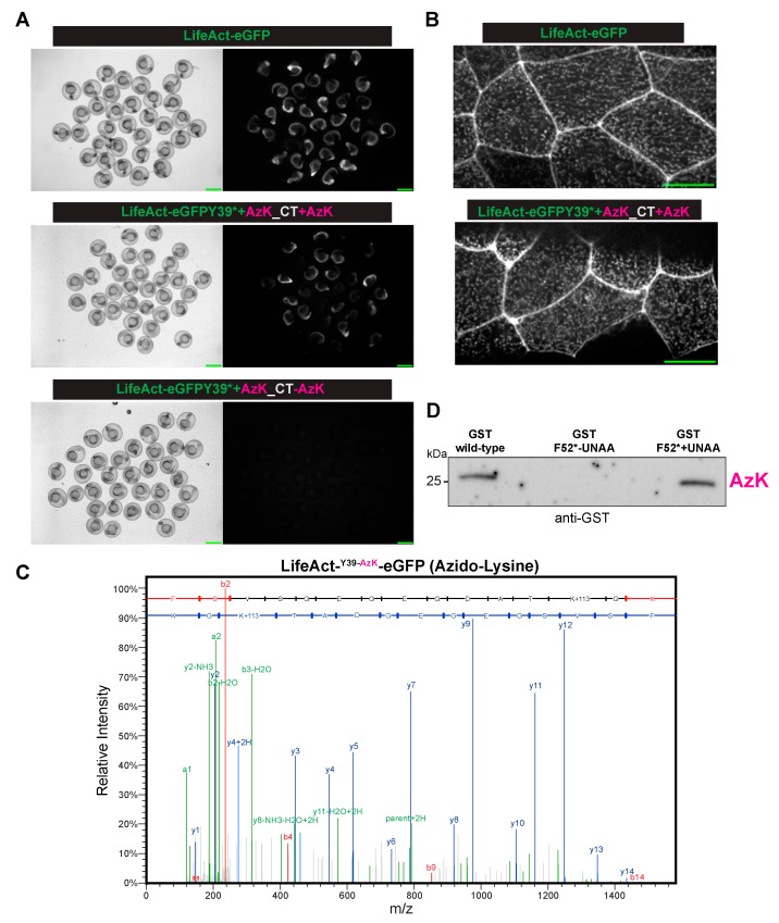
The site-specific incorporation of azido-lysine in the zebrafish embryos. (A) The Brightfield (left) or fluorescent (right) images of the zebrafish embryos injected with mRNA(s) for LifeAct-eGFP (top panel) or LifeAct-eGFP Y39* (codon for Y39 converted to stop codon UAG) with the orthogonal tRNAPyl CUA-mRNA aaRS for AzK/BCNK, with (middle panel) AzK but not without (bottom panel) AzK. Scale bar, 1 mm (B). The localization of LifeAct-eGFP or LifeAct-eGFP Y39AzK to cell–cell junctions and cortical speckles in the dechorionated embryos injected as described in (A). Scale bar represents 20 µm. (C) The mass-spectrometry identified AzK incorporation in LifeAct-eGFP Y39AzK. The lysates were prepared from ~1000 embryos injected with mRNA for LifeAct-eGFP Y39* with the orthogonal tRNAPyl-CUA -mRNA aaRS for AzK/BCNK with AzK. LifeAct-eGFP Y39AzK was immunoaffinity purified using GFP nanobodies and processed for mass spectrometry as described in the methods section. The MS/MS spectrum of precursor m/z = 791.3606 (M+2) corresponded to tryptic peptide FSVSGEGEGDATKGK with azido-lysine incorporated at the indicated position. The full y-ion fragment series is highlighted in blue. (D) An immunoprecipitation-Western blot from the zebrafish embryos injected with mRNA(s) for GST (left lane) or GST F52* (codon for F52 converted to stop codon UAG) with the orthogonal tRNAPyl-CUA -aaRS pair for AzK/BCNK, without (middle lane) or with (right lane) AzK. The immunoprecipitations and Western blots were performed with antibodies against GST.

Acknowledgments
This image is the copyrighted work of the attributed author or publisher, and ZFIN has permission only to display this image to its users. Additional permissions should be obtained from the applicable author or publisher of the image. Full text @ Int. J. Mol. Sci.