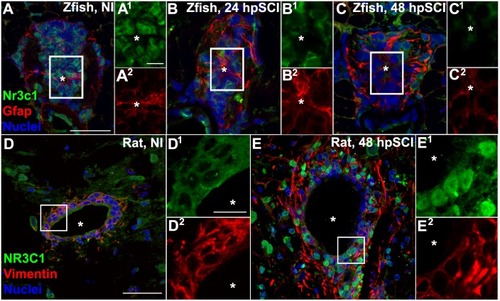
Spinal cord injury stimulates differential Nr3c1 expression by ependymal glia in zebrafish and rats. (A–C) Immunostaining for Nr3c1 (green) and Gfap (red), plus DAPI staining (blue) in larval zebrafish spinal cord transverse sections at 24 h after sham injury (A), or 24 h (B), and 48 h (C) post SCI. The nuclear Nr3c1 distribution in Gfap+-ependymal glia without injury (A1,2) is shifted to cytoplasmic at 24 and 48 h post SCI (B1,2,C1,2). Scale bars, 20 μm (A, same as B,C) and 5 μm (A1, same as A2–C2). Boxed regions denote ependymal glial cell bodies around the central canal (∗) and are magnified 167% (A1,2–C1,2). Representative images from 10 zebrafish (three sections per cord examined). See Supplementary Figure S2 for validation of Nr3c1 immunoreactivity. (D,E) Adult rat spinal cord transverse sections examined at 48 h post sham injury (D) and 48 h post SCI within 300 μm rostral of transection (E). The cytoplasmic Nr3c1 expression in ependymal glia in controls (D1,2) is shifted to the nucleus after SCI (E1,2). Scale bars, 50 μm (D, same as E) and 12.5 μm (D1, same as D2–E2). Boxed regions denote ependymal glial cell bodies around the central canal and are magnified 333% (D1,2,E1,2). Representative images from six rat spinal cords (three sections per cord examined).
|
