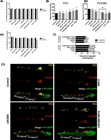
In vitro and in vivo effects of lipid nanocapsules on PC3-M8 cell viability and migration. (A) Cell viability assays performed with different TRPM8 agonists (WS12, empty LNC600, LNC600-WS12 or icilin) in PC3 and PC3-M8 cells and evaluated after 24 h (Ai) or 72 h (Aii) of incubation. MTS reagent was used to determine cell viability percentages (mean ± SEM; normalized to the control (CTRL) condition). (B) Transwell migration assays showing the effects of TRPM8 agonists on PC3 (Bi) or PC3-M8 (Bii) cells. The results are represented as a percentage of migrated cells normalized to the results observed under the CTRL condition (N = 9, mean ± SEM; *P < 0.05; **P < 0.01, ANOVA, Tukey’s multiple comparisons test for control and pairwise t-test comparison against 10 nM WS12 encapsulated or not condition, ##P < 0.01). (C) In vivo migration assays performed in zebrafish embryos. (Ci) Graph bar representing PC3 and PC3-M8 cell migration along the zebrafish tail following the application of 100 nM of empty LNC600, 100 nM of free WS12 or 100 nM of LNC600-WS12. For each condition, PC3 and PC3-M8 cells were counted and the results normalized to the total number of migrating cells (N = 15, mean ± SEM; *P < 0.05, ANOVA, Tukey’s multiple comparisons test). (Cii) Representative confocal images of Tg[Fli1-eGFP] zebrafish embryos (vasculature shown in green) injected with DiD-labeled PC3 (yellow) and DiI-labeled PC3-M8 (red) cells. Cell migration was quantified in two segments of the ventral part of the fish (1st and 2nd segments).
|
