
Fig. 5
- ID
- ZDB-FIG-190719-5
- Publication
- Teixeira Rosa et al., 2019 - Periderm invasion contributes to epithelial formation in the teleost pharynx
- Other Figures
- All Figure Page
- Back to All Figure Page
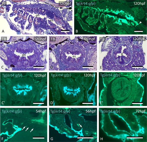
Midline krt4+ cells cover pharynx and esophagus, and meet peridermal cells invading through P3-P6. (A,B) krt4+ cells cover hyoid arch (hy) and branchial arches 1 to 5 (numbered 1–5), as well as roof and floor of the pharynx and esophagus. Note sharp boundary (arrowhead) between krt4+ esophagus and krt4- lining of the gut. (C,C’,D,D’) krt4+ cells cover the lining of the pharynx (C,C’) and the esophagus (D,D’), resp. Note long cell extensions issuing from the krt4+ cells (E,E’) Absence of krt4+ cells in the gut coincides with single-layered epithelium. (F) At 54 hpf, pouches 3–6 are still free of krt4+ cells. (G,H) Progressive invasion of P3 (arrow) by peridermal cells that meet midline krt4+ cells. (A,B,F) sagittal sections, (C–E’,G,H) cross sections. es: esophagus; g: gut; ph: pharyngeal lumen; TB: toluidine blue staining. Scale bars (A–H) = 50 μm.
|