
Fig. 2
- ID
- ZDB-FIG-190719-2
- Publication
- Teixeira Rosa et al., 2019 - Periderm invasion contributes to epithelial formation in the teleost pharynx
- Other Figures
- All Figure Page
- Back to All Figure Page
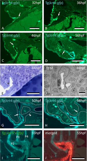
Peridermal cells sequentially invade pouches 2 to 6 but do not reach the midline endoderm. (A–C) krt4+ cells are present in progressively deeper parts of pouch 2 (P2) (arrows). (D) Peridermal cells are arrested approximately halfway the pouch (arrow). (E,F) At 44 hpf P2 is open to the exterior. Peridermal cells extend inside the pouch (black stars); a lumen forms only where these cells cover the endodermal layer. In (F), outer dotted lines mark contour of the pouch; inner dotted line marks contour of peridermal cells. (G) While peridermal cells have entered through P2 (arrow), krt4+ cells remain excluded from P3 (arrowhead) and more posterior pouches. (H) At 56 hpf, peridermal cells have started to enter P3 (arrow), and progressively extend inwards. (I,J) Double transgenics Tg(sox17:egfp;krt4:tomato) show opening of P2 with periderm entering and covering sox17+ endodermal cells (I, green channel only; J, overlay). P2–P6: pouches 2 to 6. (A–H) cross sections, (I,J) sagittal sections. TB: toluidine blue staining. Scale bars (A–E) and (G–J) = 50 μm, (F) = 5 μm.
|