
Fig. 3
- ID
- ZDB-FIG-190626-3
- Publication
- Verweij et al., 2019 - Live Tracking of Inter-organ Communication by Endogenous Exosomes In Vivo
- Other Figures
- All Figure Page
- Back to All Figure Page
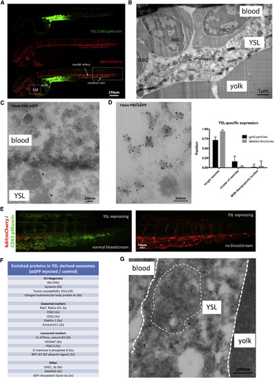
Origin and Composition of Exosomes in the Blood Flow (A, B, D, and E) Fluorescence image of a 3 dpf Tg(kdrl:Hsa.HRAS-mCherry) embryo injected with ubi:CD63-pHluorin in the YSL, green channel (top), red channel (middle), and merge (bottom). Box [EM] indicates the approximate region for EM analysis, shown in (B) and (D). Box to the right indicates the area shown in (E). (B) IEM image of the YSL of a 3 dpf Tg(kdrl:Hsa.HRAS-mCherry) embryo injected with ubi:CD63-pHluorin in the YSL labeled with gold particles directed to GFP (10 nm). Dashed white lines indicate the approximate membranes of the YSL. Dashed line box indicates area shown enlarged in (C). RBC, red blood cell. (C) Zoom in of area indicated in (B), showing MVE (asterisk) in the YSL, and EVs labeled for gold particles directed to GFP (10 nm) on top of the YSL. (D) (Left panel) IEM image of exosome-sized EVs observed in blood vessel lumen, labeled with gold particles directed to GFP (10 nm). Graph on the right: quantification of vesicle labeling and appearance as in Figure 2E (mean ± SD, >25 different fields, n = 2). (E) Zoom in on area indicated with rectangular box on the right in (A). (Left panel) Fish with blood flow; (Right panel) fish without blood flow, both expressing CD63-pHluorin in the YSL. (F) Mass-spectrometry analysis of enriched proteins in YSL-derived EVs (excerpt, complete list in Table S2) (numbers between bracket indicated the rate of enrichment in injected embryos compared to control embryos; OI, only detected in injected embryos). (G) IEM image of an MVE in the YSL area, labeled with gold particles directed to GFP (10 nm). Thin dashed white circle indicates an MVE. YSL, yolk syncytial layer; MVE, multivesicular endosome. |
Reprinted from Developmental Cell, 48(4), Verweij, F.J., Revenu, C., Arras, G., Dingli, F., Loew, D., Pegtel, M.D., Follain, G., Allio, G., Goetz, J.G., Zimmermann, P., Herbomel, P., Del Bene, F., Raposo, G., van Niel, G., Live Tracking of Inter-organ Communication by Endogenous Exosomes In Vivo, 573-589.e4, Copyright (2019) with permission from Elsevier. Full text @ Dev. Cell