
Fig. 1
- ID
- ZDB-FIG-190322-2
- Publication
- Berndt et al., 2018 - Dynamic and non-contact 3D sample rotation for microscopy
- Other Figures
- All Figure Page
- Back to All Figure Page
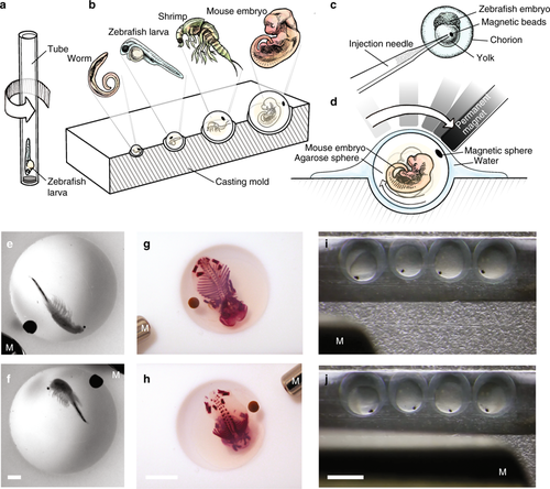
Embedding strategies for various samples and 3D non-contact orientation. a Schematic of the uniaxial rotation of a fish larva in a SPIM system. b Drawing showing the embedding of different organisms in differently sized agarose spheres by using a casting mold with hemispherical wells. c Drawing showing the injection of magnetic beads into the yolk of the zebrafish embryo. d Drawing showing a mouse embryo embedded in an agarose sphere rotated by a permanent magnet. e, f Bright-field images of a fixed artemia spec. and g, h a fixed, skeletal stained mouse embryo (E15.5) embedded in an agarose sphere and rotated by a permanent magnet (M). Scale bar, 1 mm and 5 mm, respectively. i, j Bright-field images of injected zebrafish embryos rotated by a permanent magnet. Scale bar, 1 mm |