
|
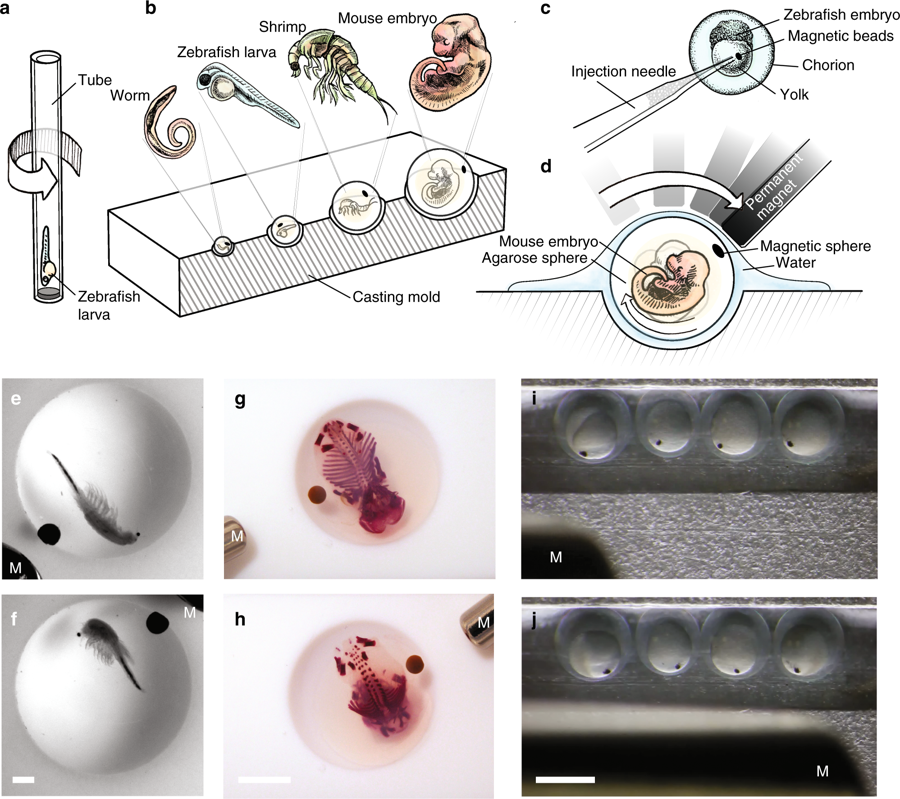
Fig. 1 Embedding strategies for various samples and 3D non-contact orientation. a Schematic of the uniaxial rotation of a fish larva in a SPIM system. b Drawing showing the embedding of different organisms in differently sized agarose spheres by using a casting mold with hemispherical wells. c Drawing showing the injection of magnetic beads into the yolk of the zebrafish embryo. d Drawing showing a mouse embryo embedded in an agarose sphere rotated by a permanent magnet. e, f Bright-field images of a fixed artemia spec. and g, h a fixed, skeletal stained mouse embryo (E15.5) embedded in an agarose sphere and rotated by a permanent magnet (M). Scale bar, 1 mm and 5 mm, respectively. i, j Bright-field images of injected zebrafish embryos rotated by a permanent magnet. Scale bar, 1 mm