
Fig. 5
- ID
- ZDB-FIG-190108-18
- Publication
- Lin et al., 2018 - Rigid Embedding of Fixed and Stained, Whole, Millimeter-Scale Specimens for Section-free 3D Histology by Micro-Computed Tomography
- Other Figures
- All Figure Page
- Back to All Figure Page
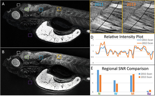
Samples embedded with our protocol can be re-imaged with micro-CT. The same 5 dpf zebrafish larva was imaged in (A) 2011 and (B) 2013, presented as average intensity projections of the sagittal view. Image comparison between scans after the storage shows the preservation of anatomical features. (C) A close-up of muscle from both scans is presented, with segmented lines indicating corresponding regions used for (D) intensity profile generation. Intensity profiles normalized to their corresponding averages along both lines show spatially matching local peaks in both scans. (E) Average intensity values for selected regions in A and B belonging to background (Bg, purple), neuronal nuclei (N, green), white matter (W, blue), and the muscle (M, orange) were divided by an average for background (white) and used to generate signal-to-noise ratios (SNR), which corresponds well between scans. Scale bars = 100 µm. |