
Fig. 4
- ID
- ZDB-FIG-190108-17
- Publication
- Lin et al., 2018 - Rigid Embedding of Fixed and Stained, Whole, Millimeter-Scale Specimens for Section-free 3D Histology by Micro-Computed Tomography
- Other Figures
- All Figure Page
- Back to All Figure Page
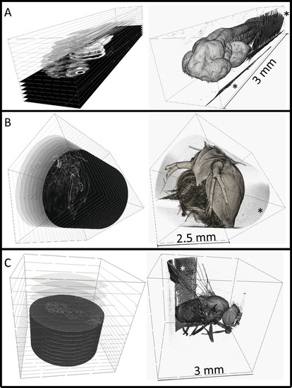
3-dimensional renderings of embedded specimens. (A) Reconstructed 3 dpf zebrafish larva at 0.743 × 0.743 × 0.743 µm3 voxel resolution. (B) Reconstructed adult Daphnia (3 × 3 × 3 µm3 voxel resolution). (C) Reconstructed adult Drosophila (3.1 × 3.1 × 3.1 µm3 voxel resolution). Digital cross-sections can be generated at any angle as shown on the left column. Surface renderings are shown on the right, rendered using commercial software. Embedding resin can be seen in all scans outside of the sample (noted by *), but does not interfere with the sample itself. The zebrafish larva was imaged at Argonne National Laboratory at the Advanced Photon Source that is synchrotron-based. The Daphnia or Drosophila specimens were imaged with a commercial X-ray microscope. |