
Fig. 3
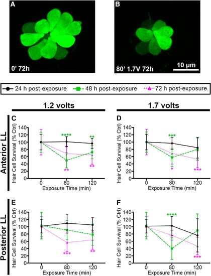
Acoustic stimulation decreases lateral line hair cell number. Unexposed (A) and acoustically stimulated (B) O2 neuromasts from myo6b:EGFP transgenic larval zebrafish. Scale bar applies to both images. 1.2 V (C) and 1.7 V (D) of acoustic stimulation significantly reduces the number of hair cells in five anterior lateral line neuromasts at 48 and 72 h after cessation of noise. 1.2 V (E) and 1.7 V (F) reduces hair cell number in pLL neuromasts P1 and P2. Asterisks indicate significant difference from age-matched unexposed controls (*p < 0.05, **p < 0.01, ***p < 0.005, ****p < 0.0001). Statistical analysis is shown in Table 1. N = 10–12 animals per treatment, values are mean ± SD. |
| Fish: | |
|---|---|
| Condition: | |
| Observed In: | |
| Stage: | Days 7-13 |