
Fig. 2
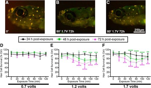
Acoustic stimulation results in exposure time-, intensity-, and post-exposure time-dependent reduction in DASPEI labeling, indicative of hair cell damage. A–C, Representative images of (A) unexposed larval zebrafish and (B, C) fish exposed to acoustic stimulation. Scale bar applies to all three images. Unexposed fish exhibit bright DASPEI staining indicative of a full complement of lateral line hair cells while fish exposed to 80 min of acoustic stimulation have diminished DASPEI labeling 72 h post-exposure. Two representative images of acoustically exposed fish are shown to depict the diversity of DASPEI labeling observed. D–F, Quantification of acoustic stimulation-induced hair cell loss. D, Fish exposed to 0.7 V show no reduction in DASPEI labeling. E, Fish exposed to 1.2 V of acoustic stimulation exhibit the greatest reduction in DASPEI labeling after 80 min of exposure and 72 h post-exposure. F, 1.7 V of acoustic stimulation produces similar DASPEI reduction to 1.2 V. Asterisks indicate significant differences from age-matched unexposed controls (*p < 0.05, **p < 0.01, ***p < 0.005, ****p < 0.001). Statistical analysis is shown in Table 1. N = 10–12 animals per treatment, values are mean ± SD. |
| Fish: | |
|---|---|
| Condition: | |
| Observed In: | |
| Stage: | Days 7-13 |